
Figure 3.
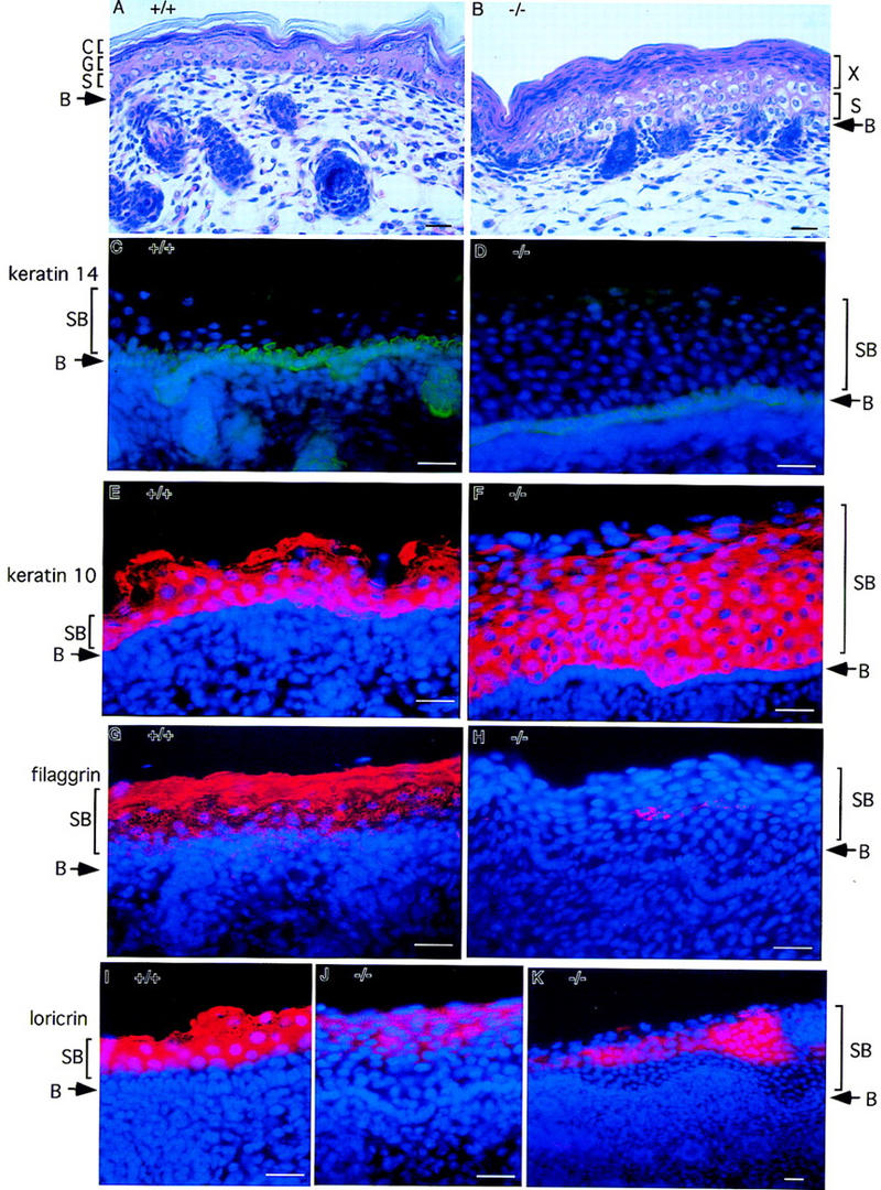
Lack of epidermal differentiation in the skin from IKK1−/− mice. Staining was performed on skin from newborn wild-type (+/+) or mutant mice (−/−). (A,B) Hematoxylin and eosin staining of skin. In the superficial layer of mutant skin, an unusual thick layer of flatten cells (layer X in B) was present instead of cornified layer. (C,D) Keratin 14 immunostaining (green) shows normal staining pattern of positive cells in the stratum basal layer. Suprabasal layer above basal layer was much thicker in mutant skin than in wild-type skin. (E,F) Anti-keratin 10 antibody stains the cells (red) in suprabasal layers in normal epidermis. The number of keratin 10 positive cells (red) in mutant skin increased dramatically. (G,H) Filaggrin is expressed in the granule and cornified layers of normal skin; however, its expression is diminished and limited to small patches of cells (red) in the mutant skin. (I–K) Expression of loricrin (red) is also reduced dramatically in the mutant skin. Nuclei (blue) were labeled by DAPI in C–K. Abbreviations: (B) Basal layer of epidermis; (S) spinous layer; (G) granular layer; (C) cornified layer; (SB) suprabasal layer. Bar size, 24 μm.