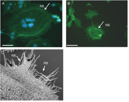
Figure 2.
(A–B) Immunofluorescence of cytoskeletal Ruffled Border (RB- white arrow); (C) SEM image of osteoclast acting ring (RB- white arrow) formed by peripheral podosome belt (P) and presenting gold immunolabelled RANKL (black arrow) at the same step of differentiation (4 days) by immunocytochemistry and SEM techniques. Scale bar: 2A–B: 50 µm. 2C 1 µm.

