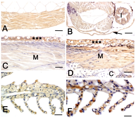
Figure 2.
Immuno-histochemical localization of IGF-I in sea bass larvae and fry. All panels are counter-stained with haematoxylin. A, C, E: diploid animals; B, D, F: triploid animals. A Sagittal section of a 6-day larva. A marked IGF-I immunostaining is present in the trunk musculature. B Transverse section of a 6-day larva. A marked IGF-I immunostaining is present in the trunk musculature, intestine (I) and skin (arrow). C–D Sagittal sections of 45-day larvae. Skin epithelium (asterisks) exhibits a marked positivity in both diploids and triploids. An immunostaining is also present in skeletal muscle (M), although in triploids (D) the reactivity is stronger than in diploids (C). Cartilage (c) is negative. E–F Gills of 60-day fry. Immunostaining is present in the epithelium of the gill filaments and the reactivity is stronger in triploids (F) than in diploids (E). Bars (A) 20 µ m, (B) 20 µm, (C) 15 µm, (D) 15 µm, (E) 12.5 µm, (F) 12.5 µm.

