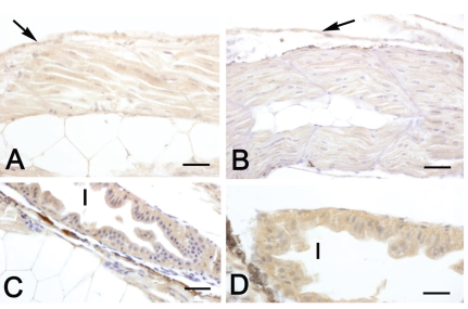
Figure 3.
Immunohistochemical localization of IGF-II in sea bass larvae. All panels are counterstained with haematoxylin. A, C: diploid animals; B, D: triploid animals. A–B Sagittal sections of 2-day larvae. In both diploids and triploids a moderate immunostaining is present in skeletal muscle and skin (arrows). C–D Sections of 10-day larvae. In both diploids and triploids a marked immunostaining is detectable in the intestinal epithelium (I). Bars (A) 15 µm; (B) 20 µm, (C) 20 µm, (D) 12.5 µm.

