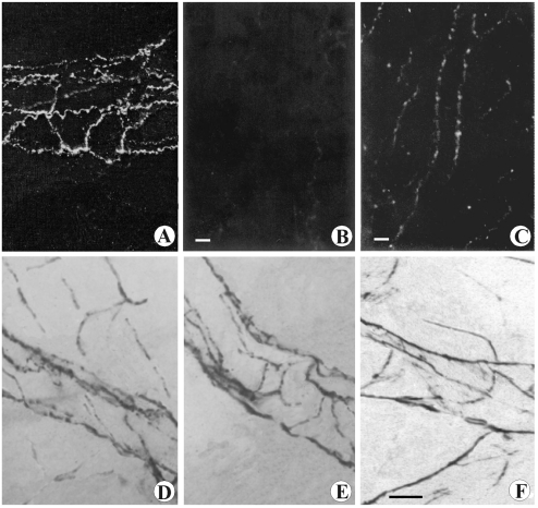
Figure 3.
Microphotograph panel showing cathecolaminergic fluorescent nerve fibers (A,B,C) and histoenzymatic AChE positive nerve fibers (D,E,F). A: Sham-operated rat, subcapsular ragion. Note the exclusive distribution of fibers forming a rich perivascular plexus. B: Surgical sympathectomised rat, subcapsular region. Note the absence of catheco-laminergic fluorescent nerve fibers. C: Sham-operated rat, medulla. Note the distribution of parallel nerve fibers running along vascular profile and several branching fibers running into parenchyma. D: Sham-operated rat, subcapsular ragion. Note the distribution of positive nerve fibers running along vessels and branching into parenchyma. E: Phrenectomised rat, subcapsular region. Note the absence of parenchymal fibers, while the AChE perivascular plexus remain well observable. F: Phrenic denerved rat, medulla. Note the occurrence of positive nerve fibers (parasympathetic fibers) running along vessels and branching into parenchyma. Calibration bar: 45 µm.

