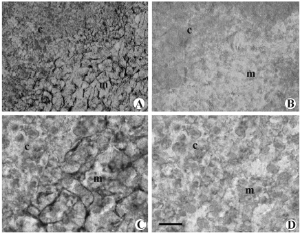
Figure 5.
Microphotograph panel of immunohistochemistry evidence of dopamine (A,B) and DAT (C,D) presence into thymus. A: Sham-operated rat. Note the occurring of positive dopamine immunohistochemistry into medullary parenchyma. B: Chemical sympathectomised rat. Note the absence of dopamine immunohistochemistry reaction. C: Sham-operated rat. Note the occurrence of positive DAT immunohistochemistry fibers into medullary parenchyma. D: Chemical sympathectomised rat. Note the absence of DAT positive reaction. c: deep cortical region; m= medulla. Calibration bar: 50 µm.

