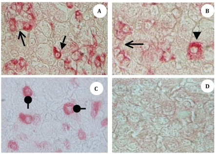
Figure 1.
A-D; Photomicrographs (X-1000) of midsagittal sections of pars distalis of moulting White Leghorn Hen (Gallus domesticus) showing (A) cephalic zone of pars distalis, ir-PRL cells (red), small arrow showing an individual ir-PRL cell with cytoplasmic extension and open head arrow is showing a islets of ir-PRL cells. Panel (B) is showing a hypertrophied cell indicated by the arrowhead; note three times bigger cell and open head arrow showing ir-PRL cells islet in crescent shape. (C) Photomicrograph of caudal lobe; roundhead arrows show ir-GH cells, noteworthy that all the cells are oval-to-round shaped with central nucleus. (D) Negative control photomicrograph.

