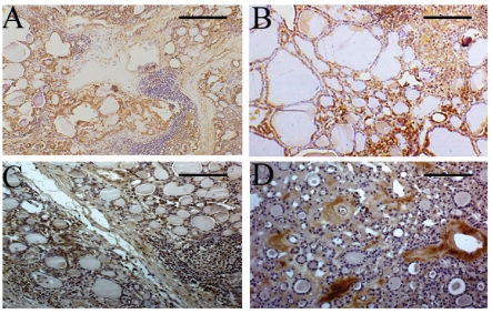
Figure 1.
HGF, c-met, PI3K and RHO immunostaining in specimens of Hashimoto's thyroditis (HT) and nodular goiters (NGs). Panel A: very intense HGF immunoreaction in a sample of HT with lymphoid aggregates defined as grade 2. The HGF immunostaining is located on the cytoplasm of thyrocytes (brown deposits) showing morphological characteristics of DN-EC cells. Note the absence of HGF reactivity in the lymphocytes (original magnification: X 150, reference bar = 133 µm). Panel B: intense c-met immunostaining in a sample of HT with grade 2 lymphoid aggregates. The c-met reaction is observed on the membrane and cytoplasm (brown deposits) of follicular DN-EC cells. No c-met reactivity is detected in lymphocytes (original magnification: X 150, reference bar = 133 µm). Panel C: moderate PI3K immunoreaction in a sample of HT with grade 2 lymphoid aggregates. The immunoreaction is located in the cytoplasm of epithelial DN-EC cells (brown deposits). Note the absence of PI3K reactivity in lymphocytes (original magnification: X 130, reference bar = 153 µm). Panel D: moderate RHO immunoreaction in a sample of NG not associated with HT. The RHO immunostaining is located on the cytoplasm of stromal cells (original magnification: X 130, reference bar = 153µm).

