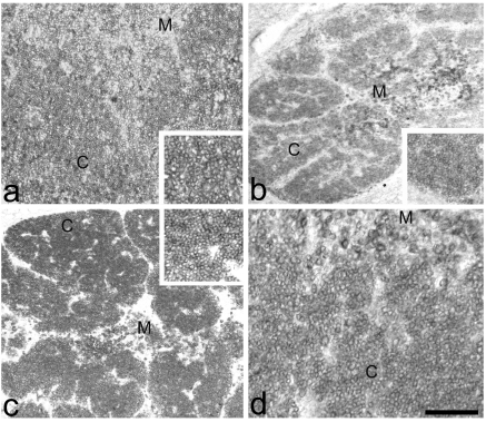
Figure 6.
Anti-CD3 immune reaction. (a) Normal thymus. Strong membrane immune staining in the cortical (C) and medullary (M) thymocytes. Inset: magnification of positive cortical thymocytes. (b) PD thymus. Reduction of the number of cortical (C) and medullary (M) reactive thymocytes. Inset: magnification of positive cortical thymocytes. (c) PD+H thymus Recovery of the total number of cortical (C) and medullary (M) reactive thymocytes. Inset: magnification of positive cortical thymocytes. (d) PD+Th thymus. Consistent recovery of the number of immune reactive thymocytes in the cortex and in the medulla. Scale bar: a: 28 µm; b,d: 40 µm; c: 50 µm.

