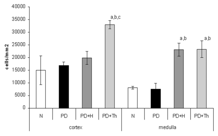
Figure 7.
Anti-CD3- Density (number of cells/mm2) of CD3+ thymocytes (detected by immune histochemistry) in thymic cortex and medulla of normal (N), partially decerebrated (PD), partially decerebrated+ hypophyseal graft (PD+H) and partially decerebrated + thymus graft (PD+Th). Data refer to quantitative analysis on tissue section and are expressed as mean ± SD. The two-tail Student t-test for unpaired data shows a significantly increase number of positive cells in cortex (a: PD+Th vs. N, P<0.001; b: PD+Th vs. PD, P<0.001; c: PD+Th vs. PD+H, P<0.001). In medulla, PD+H and PD+Th evidence a significant increment vs. N (a, P<0.001) and PD (b, P<0.001).

