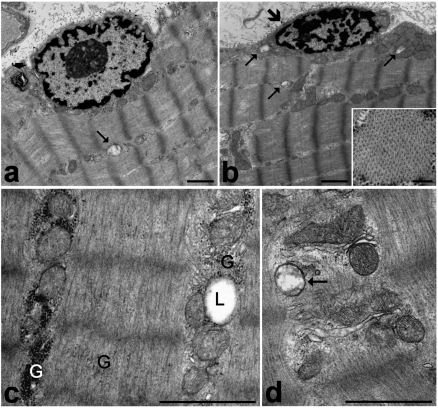
Figure 1.
Frozen muscle biopsies stored in liquid nitrogen for one (c), three (d) or four (a, b) years, then fixed with 2.5% glutaraldehyde and 2% paraformaldehyde, post-fixed with 1% OsO4 and embedded in Epon. a. The myofibre shows a well-preserved peripheral cell nucleus, cytoplasmic myofibrils longitudinally arranged in sarcomeres, and ovoid mitochondria lined along the myofibrils. Note a swollen damaged mitochondrion (arrow) and a myelin figure (arrowhead). b. A satellite cell (large arrow) showing a small heterochromatic nucleus. Arrows indicate swollen mitochondria (occurring both in the satellite cell and the adjacent myofibre) characterized by loss of cristae and a low matrix density. In the inset, a cross section of a myofibril is shown. c, d. Details of a myofibre cytoplasm. c. Glycogen (G) is present as both clusters and scattered among the myofibrils. A lipid droplet (L) is partially surrounded by mitochondria. d. The membrane structures, as well as most of the mitochondria, are well preserved and show numerous cristae and electrondense matrix granules. Note the swollen mitochondrion devoid of cristae (arrow). Bars: 1 µm; inset 0.2 µm.

