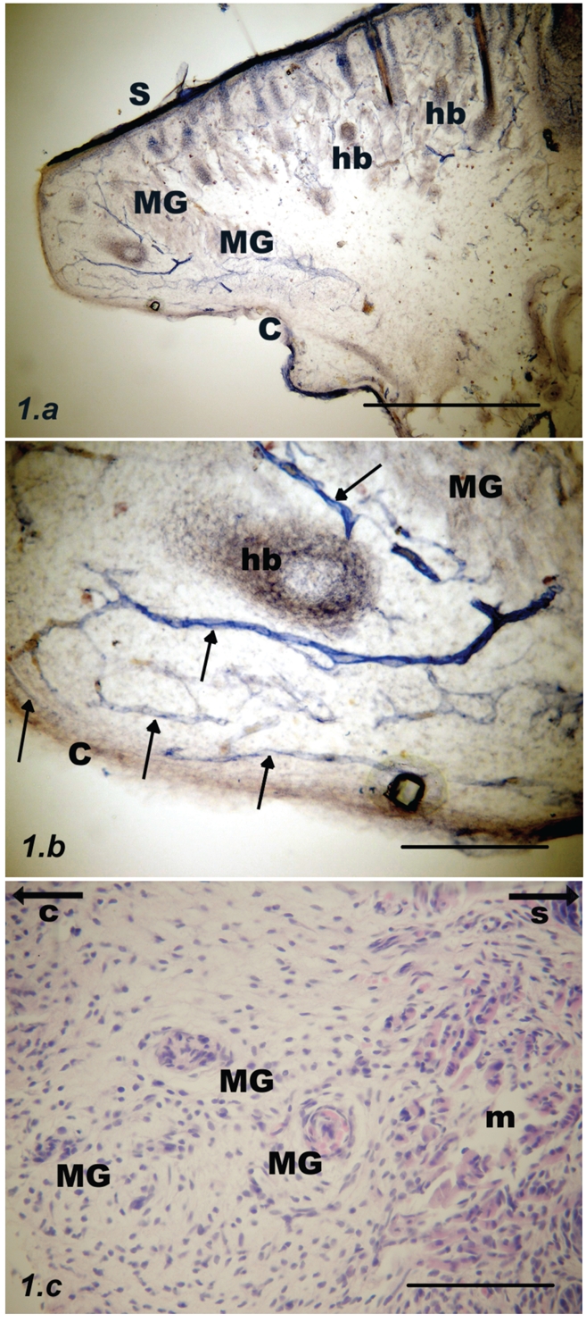
Figure 1.

Parasagittal section through the upper rat eyelid at postnatal day #1. (a) Meibomian glands (MG) are barely NADPH-d stained and are located at a distance from the tarsal conjunctival surface (C). Deep to the skin (S) there are numerous hair bulbs (hb); scale bar = 500 µm. (b) Inset from previous figure: Non-uniformly stained blood vessels (arrows) with accompanying nerve fibers. The submucous layer is relatively wide and heavily vascularised, scale bar = 100 µm. (c) Developing Meibomian glands (MG) stained with haematoxylin and eosin. Orbicularis oculi muscle fibers (m) are visible on the right. Arrows point towards the conjunctiva (C) or skin (S) respectively; scale bar = 100 µm.
