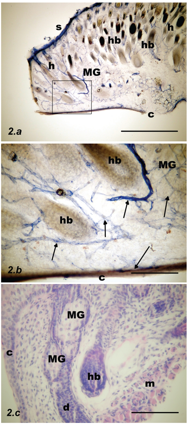
Figure 2.

Parasagittal section through the upper rat eyelid at postnatal week #1. (a) NADPH-d stain. Acini of Meibomian glands are mostly pale (MG). Deep to the skin (S) there are many hair bulbs and hairs (hb, h); scale bar = 500 µm. (b) Inset from previous figure: NADPH-d positive nerve fibers were visualised, running along the blood vessels (arrows) and in addition, single nerve fibers in the submucosa under the conjunctival epithelium (C); scale bar = 100 µm. (c) Contours of the Meibomian acini (MG) and Meibomian duct (d) shown with haematoxylin and eosin staining. A hair bulb (hb) is visible at the centre of picture and orbicularis oculi muscle fibers (m) are visible to the right; scale bar = 100 µm.
