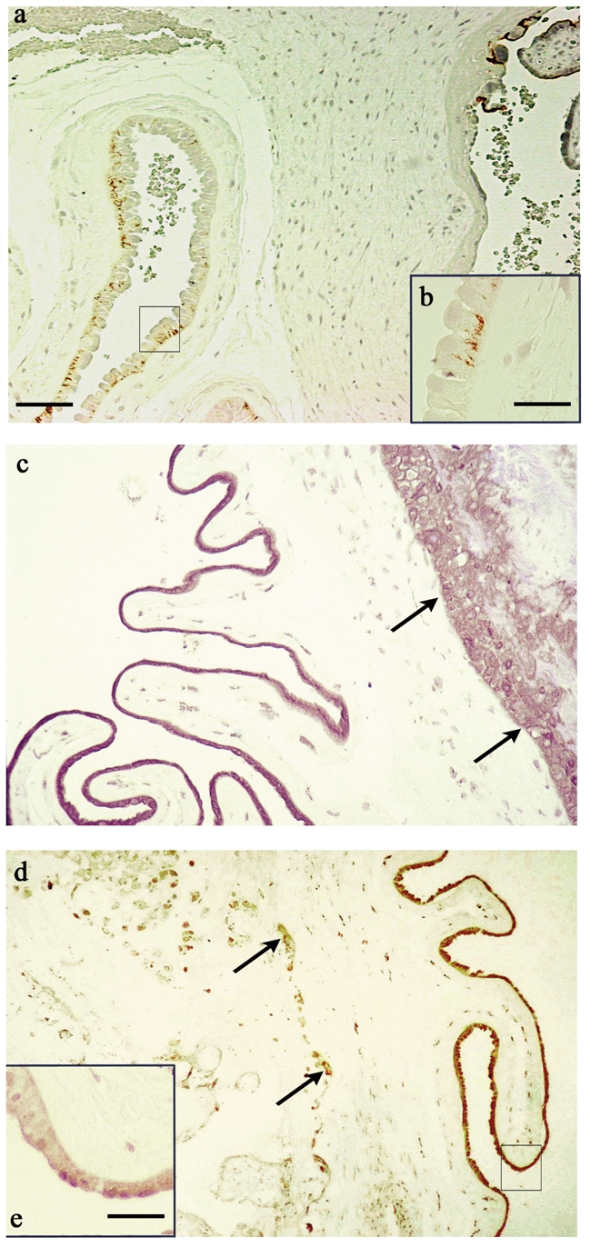
Figure 1.

Paraffin sections of term placentas. (a) Immunohistochemical localization of syndecan-1 using the antibody B-B4. (b) Shows a high magnification of the squared area depicted in panel a. Cellular plasmamembrane of amniotic epithelium shows an evident immunostaining in the basolateral compartment. (c) Immunostaining for syndecan-2 with the antibody 10H4. The amniotic epithelium and the trophoblast (arrows) underneath the amnion are positive in the cytoplasm. (d). Immunoreactivity of syndecan-4 revealed by the antibody 8G3. Arrows indicate immunostained extravillous cytotrophoblastic cells. (e) Shows a high magnification of the squared area depicted in panel (d) The staining is localized in the intracellular compartment of amniotic epithelium but it is more intense at the apex of the epithelial cells. a,c,d: Bar=90 µm; b,e: Bar=25 µm.
