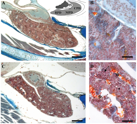
Figure 1.
Mallory stain. Sagittal sections of the P. sicula pituitary gland. (A) Control lizard showing the extension of the gland. At the top the subdivision of the gland into rostral (RPD), medial (MPD), caudal (CPD) pars distalis and pars intermedia (PI) is reported. (B) Detail of panel A showing the organization of the cellular cordons. (C) Treated lizard at 60 days. The gland tissue shows wide irregular spaces. (D) Detail of panel C: the cellular cordons show a major vascularisation (*), a basal lamina only partly surrounding them (◂) and several cells with altered shape (←). Bar 140 µm in A and C; Bar 40 µm in B and D.

