Figure 2.
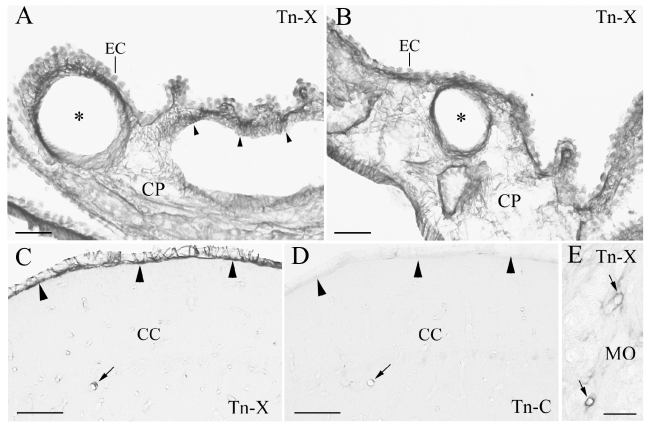
Tenascin-X (Tn-X) positive vascular and leptomenigeal structures in monkey choroid plexus (CP), cerebellar cortex (CC), and medulla oblongata (MO). (A, B) Tn-X positive fibrous structures are clearly observed around blood vessels (asterisks) and within the connective tissue of CP in lateral ventricle (arrowheads). (C) Leptomeninges on the CC is strongly Tn-X positive (arrowheads). An arrow indicates Tn-X positive blood vessel. (D) Immunohistochemistry of tenascin -C in a neighboring section to the C. Tn-C positive leptomeninges is not observed in the CC (arrowheads). An arrow indicates Tn-C positive blood vessel, which is corresponding to the Tn-X positive blood vessel in C (arrow). E: Tn-X positive blood vessels are also observed in MO (arrows). EC: epithelial cell. Scale bars=50 µm.

