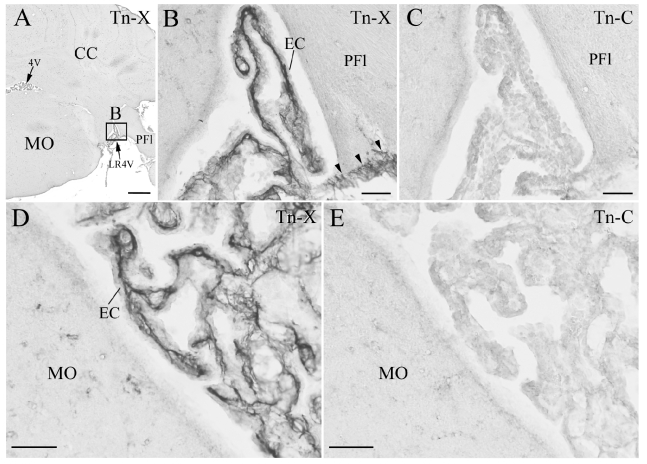
Figure 3.
Immunohistochemistry (IHC) of the tenascin-X (Tn-X) and tenascin-C (Tn-C) in mouse choroid plexus (CP) in fourth ventricle. (A) Low magnification of IHC of Tn-X in a coronal section at fourth ventricle level. One boxed region is re-photographed at higher magnification in B. (B, D) Tn-X positive fibrous structures are clearly observed within connective tissue of CP. Three arrowheads in B indicate Tn-X positive leptomeninges on PFl (paraflocculus). The panel D is photographed from an another section. (C, E) IHC of the Tn C in neighboring sections to the B and D. Tn C positive fibrous structures are not detected within the connective tissue of CP and leptomeninges. 4V: fourth ventricle, CC: cerebellar cortex, EC: epithelial cell, LR4V: lateral recess of 4V, MO: medulla oblongata. Scale bars=500 µm in A, 50 µm in B–E.

