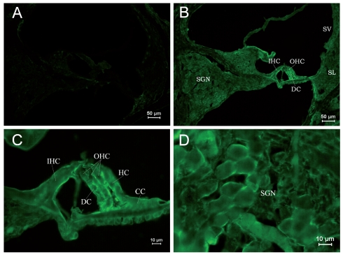
Figure 3.
Immunostain of RyRs in cochleae of the PND 10 group. (A) ×100. Negative control. Non-specific staining at the edge of the spiral lamina was weak. (B). ×100. The formation of the cochlea was basically complete. Kolliker’s organ was completely degraded. RyR expression was strong in Corti’s organ and SGNs. (C) ×400. Cells in Corti’s organ were mature. Cell arrangement was tense. IHCs, OHCs, and supporting cells were uniformly stained. (D) ×400. The staining of SGNs was similar to the PND 1 and 5 groups, but cells were not arranged in clusters. The cells were developmentally mature.

