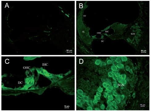
Figure 5.
Immunostaining of RyRs in cochleae of the PND 28 group. (A) ×100. Negative control. (B) ×100. Structure of mature cochleae. Strong expression of RyRs was observed in Corti’s organ and SGNs. (C) ×400. Corti’s organ was structurally mature. RyR was strongly expressed in the plasma of IHCs, especially under nuclei and near the membrane. (D) ×400. RyR expression in SGNs. RyRs were concentrated near the cell membrane.

