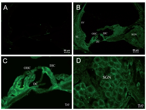
Figure 6.
Immunostaining of RyRs in cochleae of the adult group. (A) ×100. Negative control. (B) ×100. RyRs were mainly expressed in Corti’s organ. RyR staining was not complete in the plasma of SGNs. (C) ×400. The expression of RyRs was uniform in IHCs and slightly stronger at the lateral wall. The expression of RyRs was significant under the reticular lamina of OHCs and a little lower above the nuclei. (D) ×400. RyRs were expressed near the SGN membranes and were concentrated granularly in some areas.

