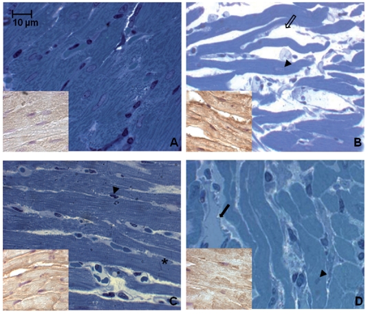
Figure 1.
Toluidine blue stained semithin sections of rat heart in different experimental conditions. Note in the hypoxic young smaller cells and increased space (thin arrow) between them occupied by abundant vessels (thick arrows) (A–B). Similar features are disclosed by the old in the two experimental conditions (cell enlargement and abundant connective compartment) (C–D). Arrowheads indicate myocardial cell nuclei. Asterisk indicates intercalated disk. Insets represent myocardial cells positivity to HIF-1 α, hypoxic condition marker. (A) normoxic young; (B) hypoxic young; (C) normoxic old; (D) hypoxic old.

