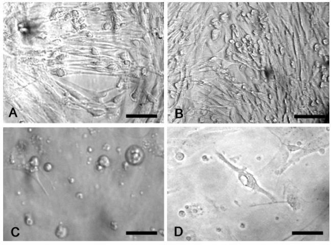
Figure 4.
Phase contrast optics. Cells exposed to 180µM 3-nitro-L-tyrosine (B) do not show any significant change in cell number and morphology in comparison with controls (A). Cells exposed to 360µM 3-nitro-L-tyrosine (C) show globular shape, short cytoplasmic processes and intracytoplasmic vacuols as a consequence of cell suffering (D). Scale bars in A, B: 80µm; C, D: 40µm.

