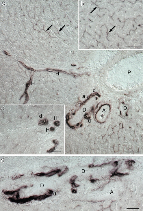
Figure 1.
Photomicrographs of histochemical demonstration of alkaline phosphatase activity in Wistar rat liver (differential interference contrast; DIC); (a) Survey view of the portal area. The reaction is very intense in small bile ducts (d), Hering's canals (H), tunica externa (adventitia) of large arteries (A) but not in arteriole (a) and in several bile canaliculi identified mostly longitudinally but also transversally (black arrows). In the lumen of one of the large arteries a positive granulocyte can be seen. P: portal vein branch. Scale bar = 50 µm; (b) Typical chicken-wire pattern of bile canaliculi (black arrows) in the portal area (see also Figure 3d). Scale bar = 50 µm; (c) Detail of AlkP activity in bile ductule (d) and Hering's canals (H) seen under higher magnification. Scale bar = 25 µm; (d) Closer view of bile ductule (d) confluence into a larger bile duct (D) Scale bar = 25 µm.

