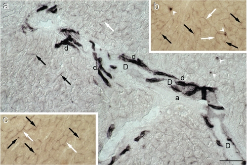
Figure 2.
Alkaline phosphatase activity in lean Zucker rat liver. (a) Survey of a portal region showing several intensely stained bile ductules (d) which abut into on a large duct (D), negative for AlkP. Moderately intense staining is present not only in bile canaliculi (black arrows) but also in basolateral (interhepatocyte) membrane domains (white arrows). The white arrow heads indicate positive sinusoidal cells. (a)=arteriole. (DIC); (b) and (c) Details of AlkP staining in interhepatocyte membrane domains (white arrows) and bile canaliculi (black arrows) (bright field) (see also Figure 3e). Scale bar = 50 µm.

