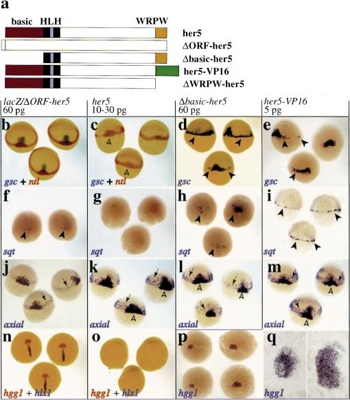
Figure 2.
her5 controls the number of cells specified to the anterior- and posterior-most mesendoderm during gastrulation. (a) Wild-type and mutant forms of her5 (red; basic DNA-binding domain; black, HLH dimerization domain; yellow, WRPW carboxy-terminal tetrapeptide; green, VP16 activation domain). (b–q) Embryos were injected at the one-cell stage with capped RNAs as indicated above each boxed area, and probed at shield-60% epiboly (b–m) or 90% epiboly (n–q) with the genes indicated on each panel (color-coded). Dorsal views, anterior to the top, except in m (side views; d, dorsal side) and q (flat-mounts of the embryos in p, anterior to the left). her5 misexpressions inhibit gsc, sqt, and hgg1 expression (c,g,o). In n–o, hlx1 labels the posterior part of the prechordal plate, so most of the prechordal plate is missing in her5-injected embryos. Conversely, Δbasic–her5 and her5–VP16 increase the number of cells expressing gsc and sqt (d,e,h,i, arrowheads). The effect of her5–VP16 perdures and induces 2.5 times more hgg1-positive cells at late gastrulation (p,q; cf. control and her5–VP16-injected embryos on the left and right of each panel, respectively). The endoderm fated to intermediate anteroposterior positions (k–m, arrows) is not significantly affected; neither are notochordal precursors [medial expression of ntl (c) and axial (k–m)] (open arrowheads; data not shown).

