Figure 6.
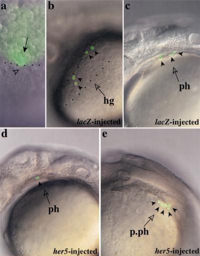
her5 expression biases cell fate choice within the endoderm/mesendoderm. (a) Fate mapping technique. Single dorsal blastomeres (arrow) of the most marginal row were labeled at 40% epiboly by uncaging photoactivatable Fluorescein DMNB with a microlaser beam (dots indicate the embryonic margin and the arrowhead points to a nucleus of the yolk syncitial layer). (b,c) Fate acquired by dorsal endomesendodermal precursors in wild-type embryos. Most progenitors contributed cells to both the hatching gland (hg) delimited by dots in b, and pharyngeal endoderm (ph) (c). Each arrowhead points to a labeled cell. (d,e) Fate acquired by dorsal endomesendodermal precursors under conditions of her5 overexpression. Most progenitors contributed to an increased number of pharyngeal (ph) or postpharyngeal (p.ph) endodermal cells only, whereas the hatching gland was populated only rarely. Two different embryos are shown, each arrowhead points to a labeled cell. Four other labeled cells are out of focus and thus not visible on the embryo in d.

