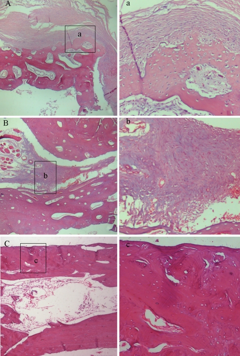
Fig. 5.
The histological microphotographs of the defects in the control group (A, a), low-dose group (B, b) and high-dose group (C, c) at eight weeks postoperatively. H-E staining; original magnification: 40× (A, B and C) and 200× (a, b and c); n = 9 per group. The defects in the control group (A) and the low-dose group (B) were filled by fibrous callus, but more vascularity and less fibroblast density appeared in the low-dose group (b), and numerous blue-violet fibroblasts were formed in the control group (a). In the dark red lamellar bone areas, no visible margin of osteotomy was seen in high-dose group, and the restored marrow cavity suggested bone remodelling (C). No fibroblast and restored blood supply were seen in c

