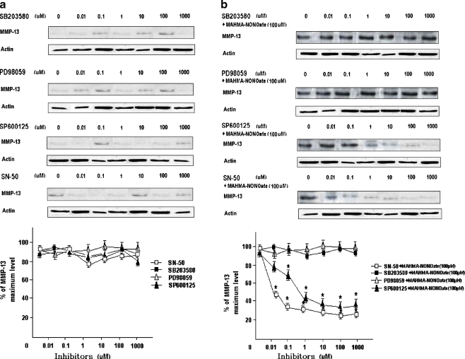
Fig. 3.
The inhibition of MMP-13 expression using the inhibitors of MAPKs and NF-κB. Cells were stimulated with various concentrations of SB203580, PD98059, SP600125 or SN-50 for 1 h respectively, and the levels of MMP-13 were determined by Western blot analysis (a). Cells were stimulated with various concentrations of SB203580, PD98059, SP600125 or SN-50 for 1 h respectively, then treated with 100 µM MAHMA-NONOate for 24 h and the levels of MMP-13 were determined by Western blot analysis (b). Points and bars are the means and standard deviations (SD), respectively, of the values obtained from three independent experiments performed in duplicate. *P < 0.05, significantly different from the control cultures

