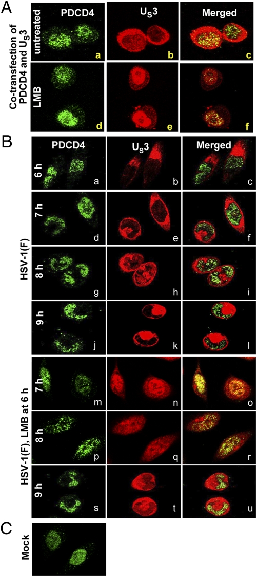
Fig. 4.
US3 shuttles between the nucleus and cytoplasm. (A) HeLa cells grown in 4-well slides were cotransfected with US3 and PDCD4 (0.1 μg of each plasmid per well). At 24 h after transfection, the medium was replaced with fresh medium of medium containing 10 μM LMB for 1 h. The cells were fixed and reacted with mouse monoclonal antibody to PDCD4 (1:200) and rabbit polyclonal antibody to US3 PK (1:1,000). (B) HeLa cells grown in 4-well slides were mock-infected or infected with 10 pfu of HSV-1(F) per cell. At 6 h after infection, the medium in a replicate set of slides was replaced with fresh medium containing 10 μM LMB. The cell cultures were fixed at 6, 7, 8, or 9 h after infection and reacted with antibodies to PDCD4 or US3 as above. For quantification of the distribution of US3 PK, digital images of adjacent fields were collected and the distribution of US3 PK within the infected cells was tabulated. The green fluorescence in A a, d and B a, d, g, j, m, p, and s shows the intracellular localization of PDCD4. The red fluorescence in A b, e, and B b, e, h, k, n, q, and t shows the intracellular localization of US3.

