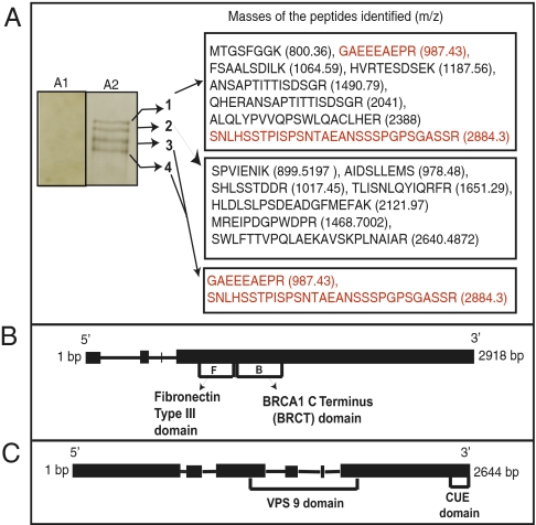
Fig. 2.
Isolation and characterization of RGD-binding and VPS9 proteins. (A1) Control sample from unmodified Sepharose column. (A2) Proteins isolated by Sepharose RGD affinity chromatography, separated by SDS/PAGE on a 10–20% gradient gel, and visualized by silver staining. Band 1 shows the amino acid sequence and mass of protein with a BRCA1 C-terminal domain and a fibronectin type III domain (red letters). Band 2 shows VPS9 with a CUE domain. Bands 3 and 4 show truncated versions of protein in band 1. (B) Gene structure of PGTG_10537.2 encoding RGD-binding protein with 818 amino acids in four exons. (C) Gene structure of PGTG_16791 encoding VPS9 with 744 amino acids.

