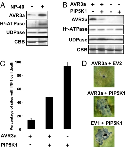
Fig. 6.
PIP5K1 OX suppresses the accumulation of AVR3a in planta. (A) Immunoblots probed with anti-FLAG antibody, anti–H+-ATPase antibody, and anti-UDPase antibody, following expression of FLAG-AVR3a in N. benthamiana. The protein was extracted with or without Nonidet P-40 (NP-40). Protein loading is shown by Coomassie blue (CBB) staining. (B) Immunoblots probed with anti-FLAG antibody, anti–H+-ATPase antibody, and anti-UDPase antibody, following expression of FLAG-AVR3a in N. benthamiana leaves overexpressing PIP5K1. (C) Percentage of sites with INF1-induced cell death upon coexpression of INF1 with AVR3a and PIP5K1. Cell death was scored 5 d after infiltration with INF1. Error bars indicate SD of the means (n = 3). (D) Symptoms observed at infiltration sites coexpressing INF1 with AVR3a and PIP5K1. A. tumefaciens strains carrying pGWB12 empty vector (EV1) or pEAQ-HT empty vector (EV2) were infiltrated as controls for AVR3a and PIP5K1, respectively.

