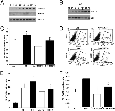
Fig. 5.
CO induces activation of DNA repair signaling. (A and B) Immunoblotting with antibody against P-Brca1 and P-ATM (A) and P-ATR (B) in the lysates of PC3 and HEK cells treated with CO (250 ppm) for 2 min to 1 h. Data are representative of three independent experiments. (C and D) U20S-SCR reporter cells were transfected with SceI for 24 h and treated with CGK733 (10 μM) or DMSO for 1 h before treatment with CO (250 ppm) for 24 h. The level of GFP+ cells was measured by flow cytometry. Quantitation and representative dot plot figures are shown in C and D, respectively. *P < 0.05, CO versus air; #P < 0.05, CO + CGK733 versus CO. (E) U20S-SCR reporter cells were cotransfected with SceI for 24 h and KU55933 (20 μM) or DMSO for 1 h before treatment with CO (250 ppm) for 24 h. The level of GFP+ cells was measured by flow cytometry. *P < 0.05, CO versus air; **P < 0.01, CO + KU55933 versus CO; (−), untransfected cells. (F) U20S-SCR reporter cells were cotransfected with SceI and HO-1 for 24 h; then CGK733 was applied for 24 h. The level of GFP+ cells was measured by flow cytometry. Data are representative of three independent experiments conducted in duplicate. *P < 0.05, HO-1 versus control (C), #P < 0.05, HO-1 + CGK733 versus HO-1.

