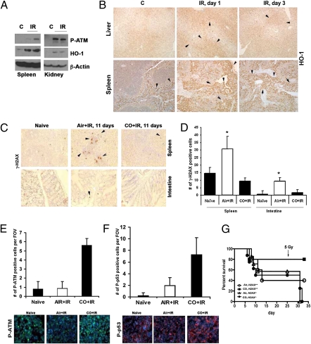
Fig. 7.
CO decreased tissue damage induced by lethal-dose irradiation. (A and B) Immunoblot (A) and immunohistochemistry (B) with antibody against HO-1 in tissues from mice irradiated with 10 Gy. (A) Kidneys and spleen harvested 2 h after irradiation. (B) Liver and spleen harvested 1 and 3 d postirradiation. (C and D) Immunohistochemical analysis of P-H2AX in the spleens and intestines of mice pretreated with air or CO (250 ppm) 1 h before a lethal dose of irradiation (10 Gy) and treated daily with CO for 11 d. Representative images are shown in C, and quantitation of the number of γ-H2AX+ cells is shown in D. n = 3 or 4 views per section from three or four mice per group. *P < 0.05. (E and F) Immunofluorescence staining of P-ATM and P-p53 in mononuclear blood cells of mice treated with CO or air for 1 h before lethal irradiation. Tissues were harvested 2 h after irradiation. Representative images (Lower) and quantitation of the number of positive cells per field of view (Upper) are shown. n = 3 or 4 mice group. (G). Survival of mice after marginal BM transplantation from H2ax−/− and H2ax+/+ mice to wild-type recipients. Mice were treated with CO before a lethal dose of irradiation (10 Gy) and after receiving BM. n = 5–10 mice per group.

