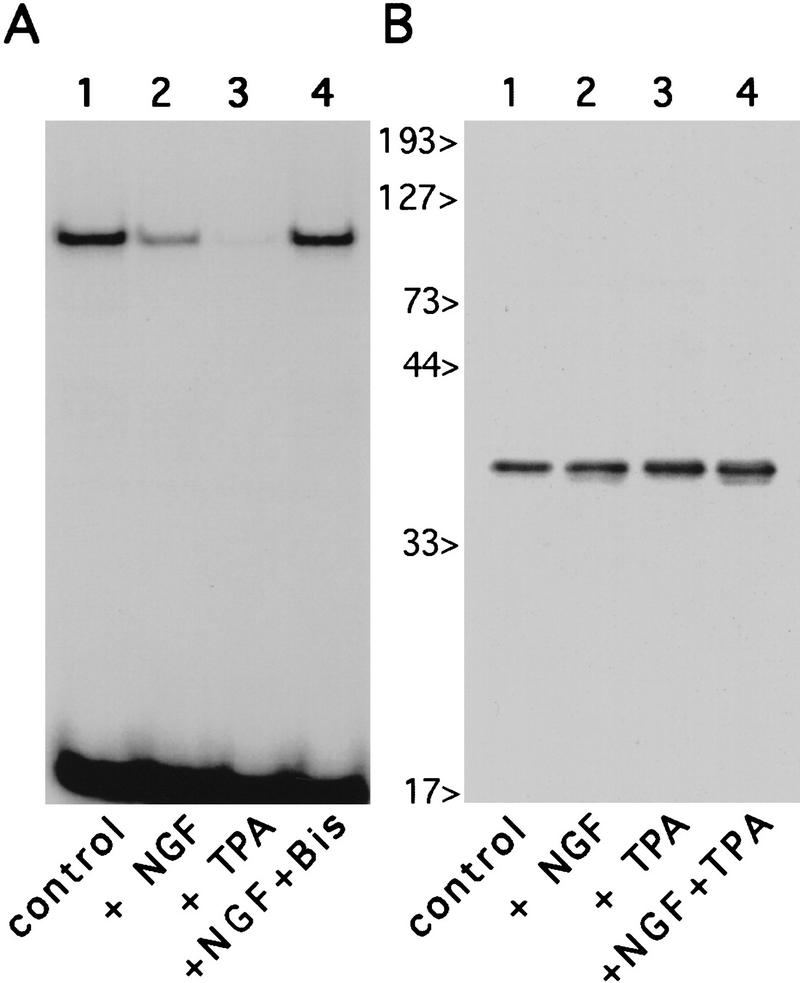
Figure 6.
Inhibition of endogenous HES-1 DNA binding during NGF- and PKC-dependent signaling. Gel-shift analysis of PC12 nuclear extracts, using a C class DNA probe, reveals that endogenous HES-1 DNA-binding activity is strongly inhibited after a 24-hr treatment with the PKC activator TPA (A, lane 3), relative to equal amounts of untreated control extract (A, lane 1). The NGF-induced decrease in HES-1 DNA binding (A, lane 2), is blocked by coaddition of the PKC inhibitor bisindolylmaleimide (A, lane 4). Western blot analysis of PC12 nuclear extracts blotted with anti-HES-1 antibody (B) reveals that, as in the case for NGF (lane 2, also see Fig. 4B), the loss of HES-1 DNA binding is not attributable to a reduction in protein levels after treatment with TPA (lane 3), or TPA plus NGF (lane 4).

