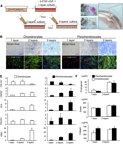
Fig. 2.
Human perichondrium-derived cells show a highly chondrogenic profile. (A) Schematic diagram of the layered culture system used for chondrogenic induction. (B) Cytochemistry staining of chondrocytes (Left) and perichondrocytes (Right) after the cells were cultured in the layered system. Perichondrocytes differentiated into chondrocytes that produced proteoglycans (Alcian blue) and type II collagen (Col2, Green). (Scale bars, 200 μm.) (C and D) Aspiration of untreated (C) and treated (D) culture media. The secretion of several mucopolysaccharides from the perichondrocytes after layered induction made the media viscous and matrix-like (Movie S1). (E) Real-time PCR analyses of gene expression profiles related to elastic cartilage in chondrocytes (Left) and perichondrocytes (Right) after the cells were cultured in the layered system. Data are shown as the mean ± SD (n = 3). (F) ELISAs specific for proteoglycans, elastic fibers (elastin), and collagen secretion from chondrocytes (Left) and perichondrocytes (Right) after the cells were cultured in monolayers or a trilayered culture. Data are shown as the mean ± SD (n = 3).

