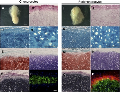
Fig. 3.
Tissue reconstitution capability of human perichondrocytes. (A and I) Chondrocytes (Left) and perichondrocytes (Right) formed cartilage-like tissues 3 mo after s.c. transplantation. (Scale bars, 1 mm.) (B and J) Hematoxylin/eosin, (C, D, K, and L) Alcian blue, (E and M) Toluidine blue, and (F and N) Safranin O staining showed that the reconstructed tissue contained mature chondrocytes in lacunae that produced high levels of proteoglycan. (G and O) Elastica Van Gieson staining showed that the reconstructed cartilage contained elastic fibers. (H and P) Immunohistochemistry revealed that perichondrocyte-derived cartilage (Right) contained type I collagen (Col1)+ capsules that enveloped a type II collagen (Col2)+ chondrium layer. (Scale bars, 200 μm.)

