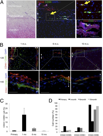
Fig. 4.
Presence and retention of CD44+ CD90+ cells in primary or regenerated perichondrium. (A) Rare population of cells in human primary auricular perichondrium (Col I+) specifically express CD44/CD90. Arrows indicate rare double positive cells existing in the outer perichondrium. (Scale bar, 50 μm.) (B) Immunohistochemical analyses of CD44 and CD90 showed apparently distinct distributions at multiple time points (1, 6, and 10 mo). Double-headed arrows show the perichondrium layers. (Scale bars, 100 μm.) (C and D) Quantification of CD44+ CD90+ cells (C) or CD44+ CD90−, CD44− CD90+, and CD44− CD90− cells (D) in primary or regenerated perichondrium.

