
Figure 4.
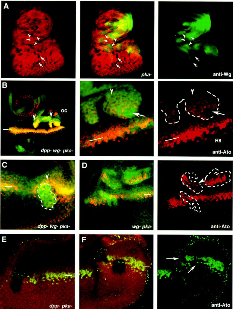
Constitutive activation of the Hh signaling pathway, in the absence of wg and dpp activities, activates the expression of the proneural gene ato and causes ectopic MFs. (A) A third instar larval eye disc stained with anti-wg (green) and carrying multiple pkaDCO–B3 clones marked by the absence of arm–lacZ (red). The arrows point to eye internal single pkaDCO–B3 clones that do not activate wg expression. The arrowheads point to clones where wg expression is activated in response to removal of pka. (B,C) Eye discs carrying dppH61 wg CX4pkaDCO–B3, which are marked by the increased accumulation of Ci occurring in the pka mutant cells (Johnson et al. 1995; M. Domínguez and E. Hafen unpubl.). The dppd12 pkaDCO–B3 clones are marked by the absence of arm–lacZ staining. (B) A large dppH61 wg CX4pkaDCO–B3 triple mutant clone (arrowheads) located in the anterior part of the eye disc. ato expression is induced autonomously in the mutant cells. A detail of the posterior part of the clone is shown in the middle panel. The right panel shows a single image of ato expression in the posterior part of the clone (outlined in white) and ato expression in the endogenous MF (indicated with the bar). In the MF (white line), the uniform levels of Ato protein evolve into discrete single Ato-positive cells, the R8 photoreceptor cells. Note that most cells in the clone have uniform levels of Ato (arrowhead) as those found in the endogenous MF. Some of the mutant cells (arrow) have initiated ommatidial development, as inferred by the presence of regularly spaced Ato-positive cells. (C) A dppH61 wg CX4pkaDCO–B3 clone located posterior to the MF at the time of dissection. The clone caused an acceleration of the endogenous MF (arrowhead). (D) Aneye disc carrying three wg CX4pkaDCO–B3 mutant clones. Note the ectopic activation of ato expression even in the clones located far from the endogenous MF. The arrowhead points to some mutant cells where ectopic expression of ato is in separated singular cells. (E,F) Two eye discs carrying dppd12 pkaDCO–B3 mutant clones. Note that ato expression (green) is not induced in these clones, which are marked by the absence of arm–lacZ staining (red). The clones in F span the endogenous MF and have caused a reduction in the levels of expression of ato (arrowheads). (oc) Occellar region. The position of the endogenous MF is indicated by white bars.