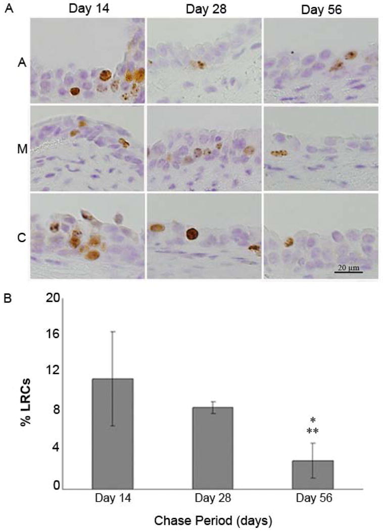
Figure 4.

A: Label-retaining cells (LRCs; brown staining) were present in basal and suprabasal layers at the anterior (A), membranous (M), and cartilaginous (C) portions of vocal fold epithelium at 14 and 28 days following injections. Minimal LRC staining was present at the A, M, and C portions of a representative vocal epithelium at 56 days post-injections. B: Mean percent label-retaining cells (%LRCs) at three points (14, 28 and 56 days post-injections). Error bars indicate ± 1 SD of the mean. * Statistical difference between day 14 and day 56 (t=4.58, p<0.05) ** Statistical difference between day 28 and day 56 (t=2.76, p<0.05).
