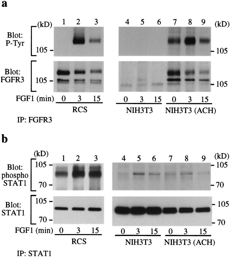
Figure 2.
FGF treatment increases STAT-1 phosphorylation in RCS cells but not in NIH-3T3 ACH cells. (a) Immunoprecipitation of FGFR3 (IP FGFR3) from RCS (lanes 1–3), NIH-3T3 (lanes 4–6), and NIH-3T3 the FGFR3 ACH mutation (lanes 7–9). The immunoprecipitates were run by SDS-PAGE and Western blotted with antibodies against phosphotyrosine (P-Tyr) (top) or FGFR3 (bottom) to determine FGFR3 expression and phosphorylation. (b) Immunoprecipitation of endogenous STAT-1 from RCS (lanes 1–3), NIH-3T3 (lanes 4–6), and NIH-3T3 ACH (lanes 7–9) cells followed by SDS-PAGE and Western blotting with antibodies specific for phosphorylated STAT-1 (top) or STAT-1 (bottom). Phosphorylation of STAT-1 in FGF-treated NIH-3T3 and in NIH-3T3 ACH cells is much weaker than in RCS cells, particularly when the total amount of STAT-1 present in the cell extracts (bottom) is considered.

