
Figure 4.
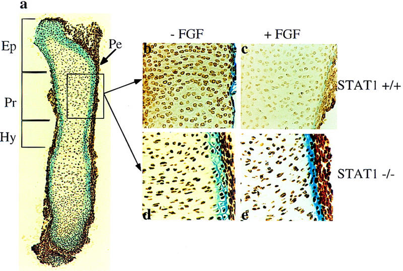
FGF inhibits chondrocyte proliferation in the epiphyseal growth plates of metatarsal bone rudiments from wild-type (STAT-1+/+) but not from STAT-1−/− mouse embryos. (a) Longitudinal section of E15 metatarsal bone rudiments after 2 days of organ culture: (Pe) Perichondrium; (Ep) epiphyseal region; (Pr) proliferative zone; (Hy) hypertrophic zone. Detailed areas of the proliferative zone stained with anti-BrdU antibody: (b,c) STAT-1+/+; (d,e) STAT-1−/−. (b,d) Untreated; (c,e) treated metatarsals with 200 ng/ml FGF1. FGF was present in the culture for 2 days, and BrdU was added for the last 6 hr of culture. Cells that incorporated BrdU were visualized as described in the text. The frequency of cells incorporating BrdU was determined by counting a total of 1000 cells in three random equal areas in the proliferative zone of three independent bone sections. In untreated STAT-1+/+ metatarsals the frequency was 76%, and in FGF-treated metatarsals, 24.7%. In STAT-1−/− metatarsals 90% of the cells had incorporated BrdU without FGF treatment, and 88% with FGF treatment.