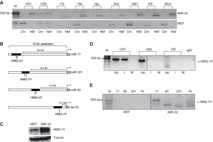
Figure 4.
Factor binding and MAR partitioning. (A) NM and Chr from IMR-32 cells and MEFs were fractionated and checked for amplification of the individual genes and the MAR elements of the miRNA 17–92 cluster. (B) Schematic representation of the putative HMG I/Y binding sites (represented by arrows) in the MAR regions (black box) of the individual miRNA genes and the miRNA 17–92 cluster (gray boxes). The distance of MARs from the each miRNA gene is also provided. (C) Immunoblot analysis of HMG I/Y in 50 µg of total lysates from MEFs and IMR-32 cells. Tubulin was used as loading control. (D) Chromatin immunoprecipitate from IMR-32 (I) or MEFs (M) were analyzed for binding of HMG I/Y protein to the upstream or downstream MAR region from miRNA 17–92 cluster. Rabbit immunoglobin served as negative control (IgG). A quantity of 10% of the total lysate was used as input (Inp). (E) HMG I/Y binding to the individual MAR elements of miRNAs 17, 93, 221 and let-7b was checked in MEFs and IMR-32 cells.

