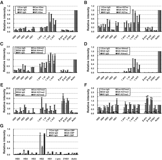
Figure 3.
Histone modification of the β-globin locus in the GATA-1 or p45/NF-E2 knockdown cells. ChIP was performed with antibodies specific to H3K9/K14ac (A), H3K27ac (B), H3K4me2 (C), H3K4me3 (D), H3K27me2 (E) and H3K27me3 (F) in K562 cells expressing each shRNA. Relative intensity was determined by normalizing to the intensity at the Actin as described in Figure 2. (G) The relative intensity of CBP ChIP was determined as described in Figure 1. Normal rabbit IgG (Con IgG) served as experimental control. The results of two to four independent experiments ± SEM are graphed.

