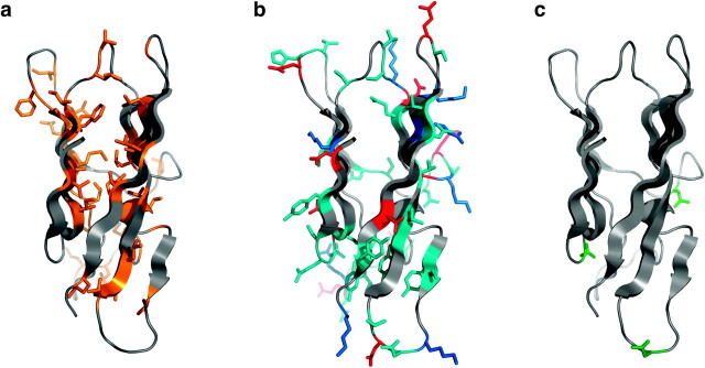
FIG. 2.
Homology model of abalone VERL repeat 10 ZP-N domain, shown in side view using a cartoon representation with relevant residues depicted as sticks. The model is consistent with burial of hydrophobic residues (a; brown), exposure of positively charged, negatively charged, and polar side chains (b; blue, red, and cyan, respectively) and exposure of consensus sites for N-glycosylation (c; green).

