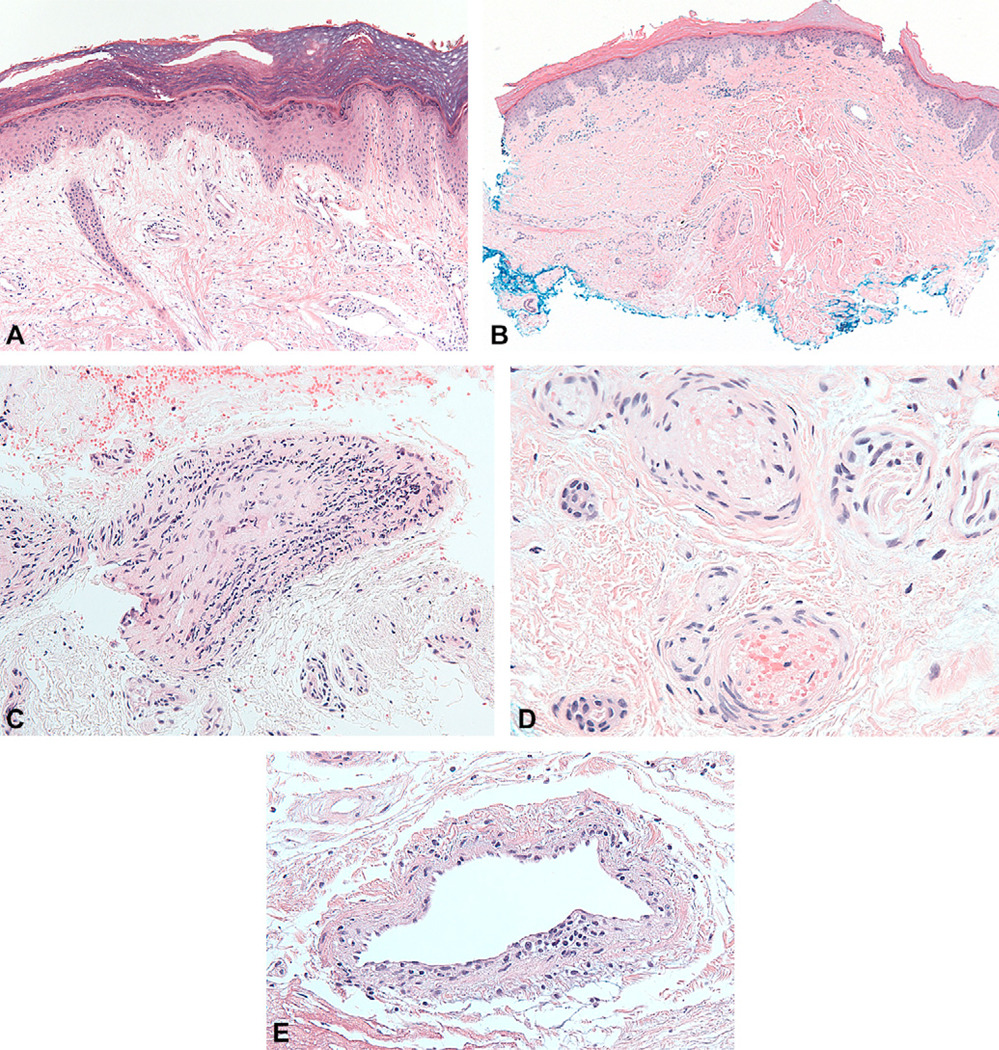
Fig 3.
Histology of palmar papules demonstrates vasculopathy; hematoxylin-eosin stain. A, Mild interface activity with increased dermal mucin. B, Pauci-inflammatory pattern in perivascular region. C, Medium vessel wall infiltration with mononuclear cells. D, Intravascular fibrin and thrombus involving small vessels. E, Endothelial cell swelling and ballooning with fibrin deposition in vessel walls.

