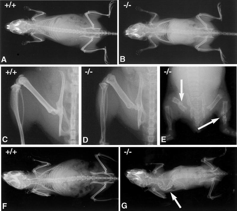
Figure 2.
Radiographic analysis of the bones of OPG−/− and wild-type mice. (A,B) Radiographs of 2-month-old female wild-type and OPG−/− mice, respectively. (C,D) Radiographs of the leg, hemipelvis and vertebrae of 2-month-old female wild-type and OPG−/− mice respectively. OPG−/− mice were x-rayed adjacent to wild-type and heterozygous mice using the same x-ray film, to allow for direct comparison of bone density. The strongest phenotype is evident in the vertebrae and long bones. The cortical bone in the femur and pelvis are thinned, and the femoral growth plate is not visible. OPG+/− mice are not different from OPG+/+ mice at this time point. (E) Gross anomalies of the skeleton are seen as early as 1 month after birth in the form of multiple fractures (arrows). (F) Radiograph of 6-month-old female wild-type mouse. (G) Radiograph of 5-month-old OPG−/− female mouse. Note severe deformity of the vertebral column due to the collapse of several vertebral bodies (arrow). This was confirmed by subsequent lateral radiographs of the spine.

