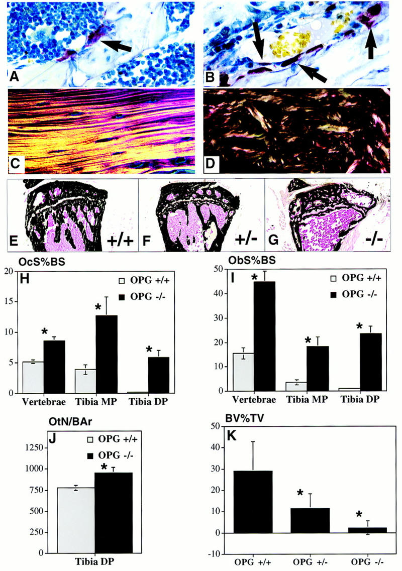
Figure 4.

Bone remodeling rate, collagen structure, and trabecular density changes in OPG−/− mice. (A,B) TRAP-stained sections (60×) of the metaphyseal region of the proximal humerus showing increased numbers of osteoclasts (arrows) in the OPG−/− (B) compared to OPG+/+ mice (A). (C,D) Polarized light microscopy of cortical bone of the humeral diaphysis showing lamellar collagen deposition in bone of the OPG+/+ (C) compared to the woven pattern in OPG−/− mice (D). (E–G) Von Kossa-stained frozen sections of the proximal tibial metaphysis of 6-month-old OPG+/+, OPG+/−, and OPG−/− mice. Note normal morphology in the OPG+/+ mice (E), lower density in the metaphysis of the OPG+/− mice (F), and even further reduced density in the OPG−/− mice (G). Osteoclast surface as a percent of bone surface (OcS%BS) (H) and osteoblast surface as a percent of bone surface (ObS%BS) (I) are increased in the vertebrae, tibial metaphysis (Tibia MP) and the tibial diaphysis (Tibia DP) of the OPG−/− (solid bars) vs. the OPG+/+ (shaded bars) mice. (*) Different from OPG+/+, P < 0.005. (J) Osteocyte number per mm2 bone area (OtN/BAr) in the tibial diaphysis is increased in OPG−/− vs. the OPG+/+ mice, P < 0.05. (K) Quantitative representation of the trabecular bone density in the metaphyseal region of the tibia expressed as a percent of the total tissue area (BV%TV) in 6-month-old OPG+/+, OPG+/−, and OPG−/− mice. The trabecular bone density was markedly reduced in the proximal tibial metaphysis in the homozygous knockout mice, n = 6, vs. heterozygous mice, n = 9, and wild type mice, n = 7. (*) Significantly lower than wild type, P < 0.001.
