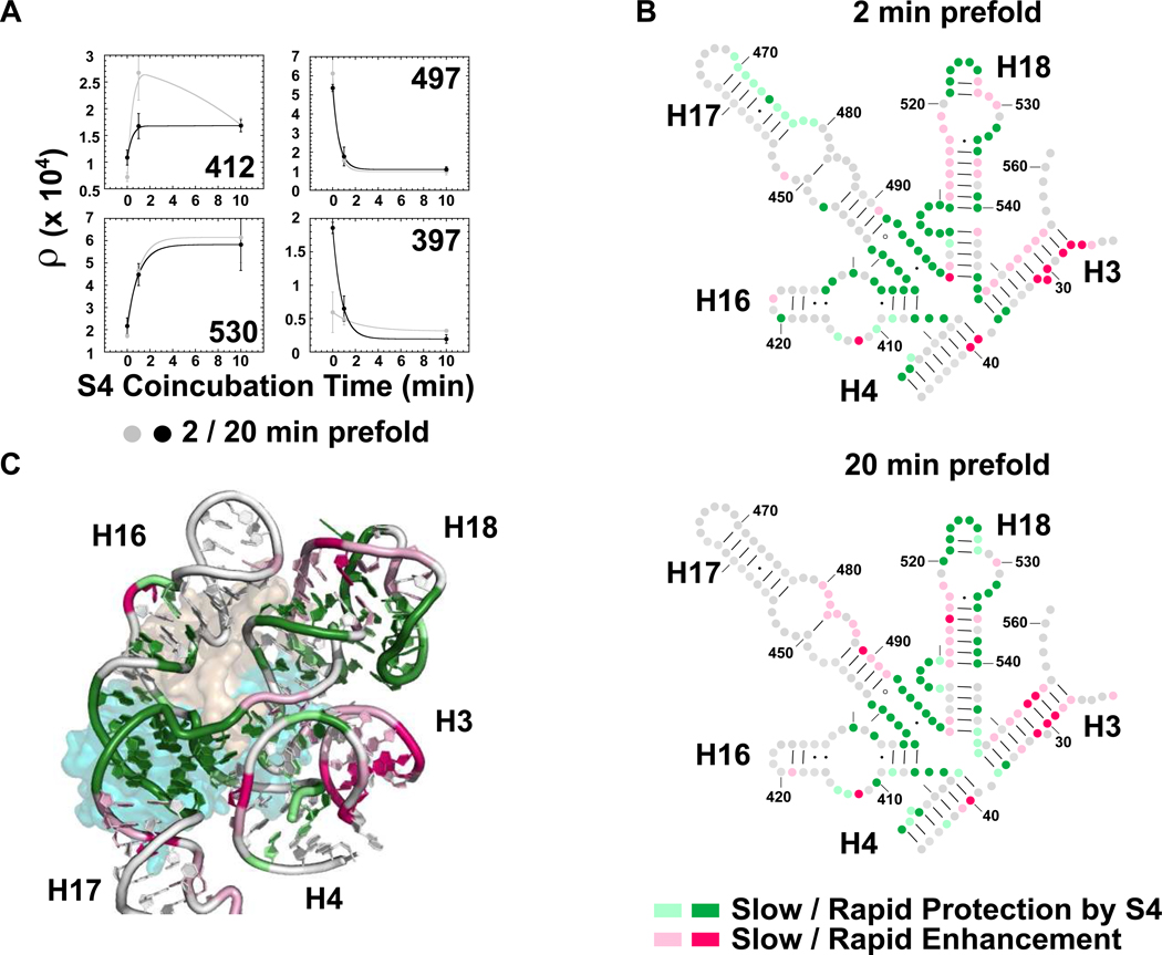
Figure 6. Time-dependence of S4-induced conformational change.
(a) SHAPE reactivity versus S4 coincubation time for four representative 5WJ nucleotides. Gray, 2 min prefold; black, 20 min prefold. Error bars as in Fig. 5a. (b) Secondary structure showing the rate of S4 protection (green) or exposure (pink) of 5WJ complexes prefolded at 42°C for 2 min (top) and 20 min (bottom). Relative differences were used to determine the % change after 1 min S4 coincubation, (ρ1min − ρ0min) / (ρ10min − ρ0min). Gray, no change; dark colors, fast (≥80% at 1 min); pale colors, slow (<80%). (c) Ribbon of E. coli S4 bound to the 5WJ in the ribosome colored as in b for 20 min prefolding (PDB:2AVY)56. S4 is colored as in Figure 1a.

