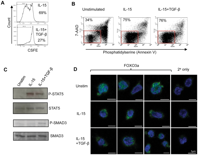
Figure 4. TGF-β does not inhibit IL-15 mediated NK cell survival.
(A) NK cell proliferation in vitro. NK cells were labeled with CFSE and cultured for five days in IL-15 alone (top panel) or IL-15 plus TGF-β (bottom panel). Cell division (as assessed by reduction in CFSE staining) was determined by flow cytometry. The percentage of cells judged to have undergone at least one cell division is shown. (B) NK cell survival in the presence or absence of IL-15. NK cells were cultured for five days in the absence of exogenous cytokines (unstimulated) or in the presence of IL-15 or IL-15 plus TGF-β. Cell survival was assessed by staining with annexin V and 7-AAD. Healthy cells (lower left quadrant) are boxed and the percentage of cells within this box are shown. (C) Signalling pathways in NK cells. Immunoblots show the phosphorylation of STAT5 (P-STAT5) and SMAD3 (P-SMAD3) in unstimulated NK cells, IL-15 stimulated or IL-15 plus TGF-β treated. Total STAT5 and SMAD3 levels are shown as loading controls. (D) Intracellular localisation of the FOXO3 transcription factor in unstimulated NK cells, NK cells stimulated with IL-15 or NK cells treated with IL-15 plus TGF-β (48 hrs) analysed by confocal microscopy. The panels show FOXO3 (in green) and the nucleus (blue) with a scale bar of 5 µm. Also shown is staining in which the anti-FOXO3 antibody was omitted and cells were stained with a secondary antibody only (2e only).

