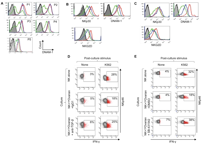
Figure 5. Human NK cell inhibition in vivo and restoration of effector function by TGF-β antagonists.
(A) DNAM-1 expression on ovarian cancer patient-derived peripheral blood NK cells (blue histograms), NK cells from the patient's ascites (green histograms) and NK cells from peripheral blood of a healthy donor (red histograms). Samples were analysed without stimulation from five patients (P1-5). NKp30, NKG2D and NKp46 expression from these five patients is shown in Figure S3. (B and C) Restoration of NK cell activation receptor expression after TGF-β antagonism in NK cell-tumour co-cultures using either an anti-TGF-β antibody in (B) or SB-431542, a small molecule inhibitor of TGF-β family receptors in (C). Primary ovarian tumour cells (from ascites fluid) were co-cultured for 48 hrs with matched peripheral blood-derived NK cells (in the presence of IL-15). The tumour cell phenotype is shown in Figure S3B. Expression of NKp30, NKG2D and DNAM-1 was analysed on NK cells cultured in the absence of matched tumour (red histograms), in the presence of TGF-β antagonists (blue histograms) or control agents (green histograms). Antagonists were the anti-TGF-β antibody in (B) or SB-431542 (C), and control agents were a control antibody in (B) or DMSO, the solvent for SB-431542, in (C). Grey filled and black histograms are isotype controls. Experiments shown in panel (B) and (C) were performed using samples from different patients. (D) and (E) Restoration of NK cell effector function by TGF-β antagonism. Patient-derived tumour cells were co-cultured with autologous NK cells (from patient peripheral blood) and IL-15 for two days in the presence of TGF-β antagonist, or control agent, as indicated. Effector function was assessed by IFN-γ production after restimulation with K562. SB-431542 also restored effector function to NK cells co-cultured with HCT116 (Figure S4). Experiments shown in (D) and (E) were performed using samples from different patients.

